lunes, 29 de abril de 2013

Characteristics of the left and right hemisphere of the brain and how they both need to be cultivated in order to attain a balanced and healthy life

Characteristics of the left and right hemisphere of the brain:

Left Hemisphere Style
Rational
  • Responds to verbal instructions
  • Problem solves by logically and sequentially looking at the parts of things
  • Looks at differences
  • Is planned and structured
  • Prefers established, certain information
  • Prefers talking and writing
  • Prefers multiple choice tests
  • Controls feelings
  • Prefers ranked authority structures
Sequential
  • Is a splitter: distinction important
  • Is logical, sees cause and effect
Draws on previously accumulated, organized information


Right Hemisphere Style
Intuitive
  • Responds to demonstrated instructions
  • Problem solves with hunches, looking for patterns and configurations
  • Looks at similarities
  • Is fluid and spontaneous
  • Prefers elusive, uncertain information
  • Prefers drawing and manipulating objects
  • Prefers open ended questions
  • Free with feelings
  • Prefers collegial authority structures
Simultaneous
  • Is a lumper: connectedness important
  • Is analogic, sees correspondences, resemblances
Draws on unbounded qualitative patterns that are not organized into sequences, but that cluster around images


What They Are, and Their Difference

Some may have heard of this, others may have not. Before we get too into specific examples to aid in our benefit, let’s go over exactly what the left and right brain are, and their associated characteristics.
Left Brain Right BrainOur brain is divided into two halves, as most of us know: the left and right side. Each side processes information very differently than the other, and the biggest difference is the visual aspect.
The right side of the brain looks at visual reference as a whole, whether it be a landscape, object, or piece of artwork, and then works its way into noticing finer details.
The left side on the other hand, first sees the details and puts them together to form the bigger picture.
Our brains use both of these sides, mixing and matching each side’s abilities for a fully-functional human brain. However, each of us has a dominant side that leans more towards the behaviors of that respected side.

Their Relation to Art, Design, and Creativity

As one may have probably already guessed, those with dominance in the right brain may be more naturally creative.
It’s easy to assume this because for one, right-brained thinkers are less common than left, so it seems as though one would be seeing the world differently from everyone else.
Also, the natural heightened visual nature and curiosity tend to make the mind never stop thinking of the alternative — as well as how it can be applied visually.
Those with a dominant left brain are far more common, and far more analytical. They may feel at disadvantage for not having that ‘natural’ creativity. Realistically, though, left-brained people can be just as creative; they just come about it in a different way.
To better understand the artistic nature of both sides, let’s take a look at a few examples of artwork.

Benefit Your Work

Let’s now look into some further ideas of how we can specifically better our work by understanding our own psychology.
After taking the tests above, you may have found out that we are not either 100% right brained, or 100% left brained. We are a mix of them both, while some traits may lean far to the opposite side, and other may not.
Also, in certain traits, we may only be a certain percentage right/left brained, while the remainder of the percentage leads the opposite way.
We each have such unique characteristics, and an in-depth analysis of each (second test listed above) can help.


viernes, 19 de abril de 2013

the different types of brain traumas and how can be treated

TRAUMATIC BRAIN INJURY:
Traumatic brain injury (TBI), also known as intracranial injury, occurs when an external force traumatically injures the brain. TBI can be classified based on severity, mechanism (closed or penetrating head injury), or other features (e.g., occurring in a specific location or over a widespread area). Head injury usually refers to TBI, but is a broader category because it can involve damage to structures other than the brain, such as the scalp and skull.
TBI is a major cause of death and disability worldwide, especially in children and young adults. Males sustain traumatic brain injuries more frequently than do females. Causes include falls, vehicle accidents, and violence. Prevention measures include use of technology to protect those suffering from automobile accidents, such as seat belts and sports or motorcycle helmets, as well as efforts to reduce the number of automobile accidents, such as safety education programs and enforcement of traffic laws.
Brain trauma can be caused by a direct impact or by acceleration alone. In addition to the damage caused at the moment of injury, brain trauma causes secondary injury, a variety of events that take place in the minutes and days following the injury. These processes, which include alterations in cerebral blood flow and the pressure within the skulL, contribute substantially to the damage from the initial injury.
TBI can cause a host of physical, cognitive, social, emotional, and behavioral effects, and outcome can range from complete recovery to permanent disability or death. The 20th century saw critical developments in diagnosis and treatment that decreased death rates and improved outcome. Some of the current imaging techniques used for diagnosis and treatment include CT scans computed tomography and MRIs magnetic resonance imaging. Depending on the injury, treatment required may be minimal or may include interventions such as medications, emergency surgery or surgery years later. Physical therapy, speech therapy, recreation therapy, occupational therapy and vision therapy may be employed for rehabilitation.

HOW CAN BE TREATED:
It is important to begin emergency treatment within the so-called "golden hour" following the injury. People with moderate to severe injuries are likely to receive treatment in an intensive care unit followed by a neurosurgical ward. Treatment depends on the recovery stage of the patient. In the acute stage the primary aim of the medical personnel is to stabilize the patient and focus on preventing further injury because little can be done to reverse the initial damage caused by trauma. Rehabilitation is the main treatment for the subacute and chronic stages of recovery. International clinical guidelines have been proposed with the aim of guiding decisions in TBI treatment, as defined by an authoritative examination of current evidence.

Acute stage

Certain facilities are equipped to handle TBI better than others; initial measures include transporting patients to an appropriate treatment center. Both during transport and in hospital the primary concerns are ensuring proper oxygen supply, maintaining adequate cerebral blood flow, and controlling raised intracranial pressure (ICP), since high ICP deprives the brain of badly needed blood flow and can cause deadly brain herniation. Other methods to prevent damage include management of other injuries and prevention of seizures.
Neuroimaging is helpful but not flawless in detecting raised ICP. A more accurate way to measure ICP is to place a catheter into a ventricle of the brain, which has the added benefit of allowing cerebrospinal fluid to drain, releasing pressure in the skull. Treatment of raised ICP may be as simple as tilting the patient's bed and straightening the head to promote blood flow through the veins of the neck. Sedatives, analgesics and paralytic agents are often used. Hypertonic saline can improve ICP by reducing the amount of cerebral water (swelling), though it is used with caution to avoid electrolyte imbalances or heart failure. Mannitel, an osmotic diuretic, was also studied for this purpose, but such studies have been heavily questioned. Diuretics, drugs that increase urine output to reduce excessive fluid in the system, may be used to treat high intracranial pressures, but may cause hypovolemia (insufficient blood volume). Hyperventilation (larger and/or faster breaths) reduces carbon dioxide levels and causes blood vessels to constrict; this decreases blood flow to the brain and reduces ICP, but it potentially causes ischemia and is, therefore, used only in the short term.
Endotracheal intubation and mechanical ventilation may be used to ensure proper oxygen supply and provide a secure airway. Hypotension (low blood pressure), which has a devastating outcome in TBI, can be prevented by giving intravenous fluids to maintain a normal blood pressure. Failing to maintain blood pressure can result in inadequate blood flow to the brain. Blood pressure may be kept at an artificially high level under controlled conditions by infusion of norepinephrine or similar drugs; this helps maintain cerebral perfusion. Body temperature is carefully regulated because increased temperature raises the brain's metabolic needs, potentially depriving it of nutrients. Seizures are common. While they can be treated with benzodiazepines, these drugs are used carefully because they can depress breathing and lower blood pressure. TBI patients are more susceptible to side effects and may react adversely or be inordinately sensitive to some pharmacological agents. During treatment monitoring continues for signs of deterioration such as a decreasing level of consciousness.
Traumatic brain injury may cause a range of serious coincidental complications which include cardiac arrhythmias and neurogenic pulmonary edema. These conditions must be adequately treated and stabilised as part of the core care for these patients.
Surgery can be performed on mass lesions or to eliminate objects that have penetrated the brain. Mass lesions such as contusions or hematomas causing a significant mass effect (shift of intracranial structures) are considered emergencies and are removed surgically. For intracranial hematomas, the collected blood may be removed using suction or forceps or it may be floated off with water. Surgeons look for hemorrhaging blood vessels and seek to control bleeding. In penetrating brain injury, damaged tissue is surgically debrided, and craniotomy may be needed. Craniotomy, in which part of the skull is removed, may be needed to remove pieces of fractured skull or objects embedded in the brain. Decompressive craniectomy (DC) is performed routinely in the very short period following TBI during operations to treat hematomas; part of the skull is removed temporarily (primary DC DC performed hours or days after TBI in order to control high intracranial pressures (secondary DC) has not been shown to improve outcome in some trials and may be associated with severe side effects.

martes, 16 de abril de 2013

Types of brain traumas and how they can be treated

Types of Brain Injury

All brain injuries are unique.  The brain can receive several different types of injuries depending on the type of force and amount of force that impacts the head. The type of injury the brain receives may affect just one functional area of the brain, various areas, or all areas of the brain.

Traumatic Brain Injury

Even a concussion can cause substantial difficulties or impairments that can last a lifetime. Whiplash can result in the same difficulties as head injury. Such impairments can be helped by rehabilitation, however many individuals are released from treatment without referrals to brain injury rehabilitation, or guidance of any sort.
  • A concussion can be caused by direct blows to the head, gunshot wounds, violent shaking of the head, or force from a whiplash type injury.
  • Both closed and open head injuries can produce a concussion. A concussion is the most common type of traumatic brain injury.
  • A concussion is caused when the brain receives trauma from an impact or a sudden momentum or movement change. The blood vessels in the brain may stretch and cranial nerves may be damaged.
  • A person may or may not experience a brief loss of consciousness.
  • A person may remain conscious, but feel dazed.
  • A concussion may or may not show up on a diagnostic imaging test, such as a CAT Scan.
  • Skull fracture, brain bleeding, or swelling may or may not be present. Therefore, concussion is sometimes defined by exclusion and is considered a complex neurobehavioral syndrome.
  • A concussion can cause diffuse axonal type injury resulting in temporary or permanent damage.
  • A blood clot in the brain can occur occasionally and be fatal.
  • It may take a few months to a few years for a concussion to heal

Acquired Brain Injury

Acquired Brain Injury, (ABI), results from damage to the brain caused by strokes, tumors, anoxia, hypoxia, toxins, degenerative diseases, near drowning and/or other conditions not necessarily caused by an external force.

Anoxia

Anoxic Brain Injury occurs when the brain does not receive any oxygen. Cells in the brain need oxygen to survive and function.
Types of Anoxic Brain Injury
  • Anoxic Anoxia- Brain injury from no oxygen supplied to the brain
  • Anemic Anoxia- Brain injury from blood that does not carry enough oxygen
  • Toxic Anoxia- Brain injury from toxins or metabolites that block oxygen in the blood from being used Zasler, N. Brain Injury Source, Volume 3, Issue 3, Ask the Doctor

Hypoxic

A Hypoxic Brain Injury results when the brain receives some, but not enough oxygen.
Types of Hypoxic Brain Injury
  • Hypoxic Ischemic Brain Injury, also called Stagnant Hypoxia or Ischemic Insult- Brain injury occurs because of a lack of blood flow to the brain because of a critical reduction in blood flow or blood pressure.

viernes, 12 de abril de 2013

The Brain functions

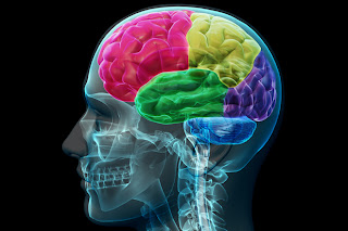
The Cerebrum: The cerebrum or cortex is the largest part of the human brain, associated with higher brain function such as thought and action. The cerebral cortex is divided into four sections, called "lobes": the frontal lobe, parietal lobe, occipital lobe, and temporal lobe.
What do each of these lobes do?
  • Frontal Lobe- associated with reasoning, planning, parts of speech, movement, emotions, and problem solving
  • Parietal Lobe- associated with movement, orientation, recognition, perception of stimuli
  • Occipital Lobe- associated with visual processing
  • Temporal Lobe- associated with perception and recognition of auditory stimuli, memory, and speech

The Cerebellum: The cerebellum, or "little brain", is similar to the cerebrum in that it has two hemispheres and has a highly folded surface or cortex. This structure is associated with regulation and coordination of movement, posture, and balance. The cerebellum is assumed to be much older than the cerebrum, evolutionarily. What do I mean by this? In other words, animals which scientists assume to have evolved prior to humans, for example reptiles, do have developed cerebellums. However, reptiles do not have neocortex. Go here for more discussion of the neocortex or go to the following web site for a more detailed look at evolution of brain structures and intelligence: "Ask the Experts": Evolution and Intelligence Limbic System: The limbic system, often referred to as the "emotional brain", is found buried within the cerebrum. Like the cerebellum, evolutionarily the structure is rather old. This system contains the thalamus, hypothalamus, amygdala, and hippocampus.
Brain Stem: Underneath the limbic system is the brain stem. This structure is responsible for basic vital life functions such as breathing, heartbeat, and blood pressure. Scientists say that this is the "simplest" part of human brains because animals' entire brains, such as reptiles (who appear early on the evolutionary scale) resemble our brain stem.    The brain stem is made of the midbrain, pons, and medulla.

Thalamus: Thalamus a large mass of gray matter deeply situated in the forebrain at the topmost portion of the diencephalon. The structure has sensory and motor functions. Almost all sensory information enters this structure where neurons send that information to the overlying cortex. Axons from every sensory system (except olfaction) synapse here as the last relay site before the information reaches the cerebral cortex.

Hypotalamus: Hypothalamus part of the diencephalon, ventral to the thalamus. The structure is involved in functions including homeostasis, emotion, thirst, hunger, circadian rhythms, and control of the autonomic nervous system. In addition, it controls the pituitary.

Amygadala: Amygdala part of the telencephalon, located in the temporal lobe; involved in memory, emotion, and fear. The amygdala is both large and just beneath the surface of the front, medial part of the temporal lobe where it causes the bulge on the surface called the uncus. This is a component of the limbic system.

Hippocampus: Hippocampus the portion of the cerebral hemisphers in basal medial part of the temporal lobe. This part of the brain is important for learning and memory for converting short term memory to more permanent memory, and for recalling spatial relationships in the world about us.

Midbrain: Midbrain Mesencephalon- the rostral part of the brain stem, which includes the tectum and tegmentum. It is involved in functions such as vision, hearing, eyemovement, and body movement. The anterior part has the cerebral peduncle, which is a huge bundle of axons traveling from the cerebral cortex through the brain stem and these fibers (along with other structures) are important for voluntary motor function.

Pons: Pons part of the metencephalon in the hindbrain. It is involved in motor control and sensory analysis... for example, information from the ear first enters the brain in the pons. It has parts that are important for the level of consciousness and for sleep. Some structures within the pons are linked to the cerebellum, thus are involved in movement and posture.

 Medulla: Medulla Oblongata this structure is the caudal-most part of the brain stem, between the pons and spinal cord. It is responsible for maintaining vital body functions, such as breathing and heartrate